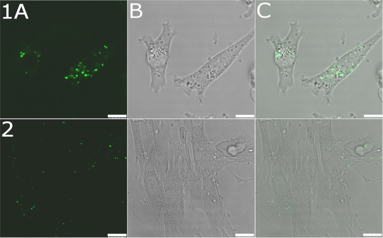
Figure 2. Confocal microphotographs of MDA-MB-231 and MRC-5 pd30 cells.
MDA-MB-231 cells (upper panel 1) and MRC-5 pd30 (bottom panel 2) were treated for 5 h with the OLICARB1:1 nanoparticles fluorescently labeled by encapsulation of 5-carboxyfluorescein; the final concentration of platinum and olaparib in the CF-labelled OLICARB nanocapsules was 1 µM, and that of CF was 0.1 µM. (A) Fluorescence channel. (B) Bright field. (C) Overlay of the fluorescence and bright field channels. Scale bar represents 10 μm for panel 1 and 25 μm for panel 2.

