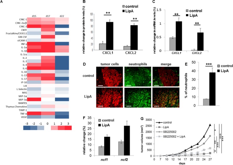
Figure 2. Anti-tumor effect of LipA was correlated with neutrophil infiltration.
(A) The modulation of cytokine and chemokine contents of tumors one day after the first, second and fourth injections (respectively d15, d17 and d22) of LipA or physiological solution was analyzed using antibody arrays and Image J software. (B) The relative abundance of CXCL1 and CXCL2 proteins twenty-four hours after the second injection of LipA, (d17) was expressed as a percentage compared to an internal control (100%). (C) Six hours after the first injection (d15) of LipA or physiological solution (control), expression levels of cxcl1 and cxcl2 mRNA in tumors were evaluated relative to housekeeping gene gapdh by RT-PCR. (D) Tumors were collected at day 17 from LipA treated or control rats. After fixation, 5-µm sections were cut and stained for tumor cells (anti-cytokeratin Ab, red) and neutrophils (anti-HIS48 Ab, green) (scale bars = 50 µm). The yellow staining in the merge panels represents tumor cells very close to neutrophils. Micrographs are representative of at least 3 independent experiments, 4 animals per group. (E) The levels of neutrophils present in tumors were determined by counting of these cells in 3 independent slides per animals, 4 animals per group. Shown are the mean % of neutrophils ± SEM. (F) At day 17, expression levels of ncf1 and ncf2 mRNA were evaluated relative to housekeeping gene gapdh by RT-PCR. (G) CT26-bearing mice were treated with physiological solution (control) or LipA with or without the GZMB inhibitor SB225002. Three days after cell injection, LipA was administrated i.v. every 5 days for 5 times. SB225002 was injected i.p. 24 h before and at the same time than LipA. Results are representative of at least 2 independent experiments with 10 animals per group. (B, C, E–G) Significant difference in Mann–Whitney U test, *p < 0.05, **p < 0.01, ***p < 0.005.

