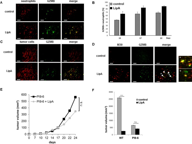
Figure 4. Granzyme B was present in neutrophils and apoptotic tumor cells in LipA treated rats.
Tumors from rats were removed at day 17, fixed, cut into 5-μm cryosections and stained. (A) Tumor associated-neutrophils containing GZMB were double stained (Neutrophils in red, GZMB in green). (B) The levels of neutrophils containing GZMB were determined at days 15, 17 and 22 by counting of these cells in 3 independent slides per animals, 4 animals per group. Shown are the mean % of double positive cells ± SEM. (C, D) Tumor sections were stained for tumor cells (red (C)), or apoptotic tumor cells (anti-M30 Ab, red (D)) and GZMB (anti-GZMB Ab, green (C, D)). Enlargement of regions a and b in figure (D) Micrographs are representative of at least 3 independent experiments, 4 animals per group (scale bars = 50 µm). (E, F) Three days after s.c. injection of PI9-6 cells (E, F) or CT26 wild type cells (WT, F), mice were treated i.v. with LipA or physiological solution (control) every 5 days for 5 times. Results are representative of at least 2 independent experiments with 10 animals per group. Significant difference in Mann–Whitney U test, ***p < 0.005, n.s. not significant.

