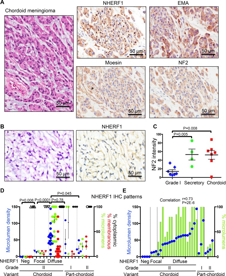
Figure 3. NHERF1, moesin and NF2 expression in chordoid meningioma.
(A) IHC with NHERF1, moesin, NF2 and EMA antibodies in chordoid meningioma shows labeling of microlumens by NHERF1 and to a lower extent by moesin and EMA. NF2 displays strong cytoplasmic expression. (B) In a minority of chordoid meningioma cases, NHERF1 expression is limited to the cytoplasm only. (C) Quantification of NF2 expression in secretory (n = 4) and chordoid (n = 7) meningioma as described in Materials and Methods, in comparison to WHO grade I meningioma cases with common meningothelial (n = 3), transitional (n = 2), fibrous (n = 1), and psammomatous (n = 1) morphologies. All the samples were run in one experiment to allow comparison. This staining was repeated three times on additional samples and showed similar results. A positive control represented by choroid plexus papilloma [8] was included in each series. (D) NHERF1 expression patterns in chordoid and part-chordoid meningioma cases represented as mean ± SEM of individual values. Each value corresponds to a case. The microlumen density, expressed as mean of counts in 3 HPFs, is plotted on the left Y axis, and the tumor areas of the three patterns—microlumens, membranous, cytoplasmic—expressed in %, are plotted on the right Y axis. Open and filled symbols for the cytoplasmic pattern correspond to weak or strong NHERF1 expression, respectively. (E) Contingency graph showing significant correlation between microlumen density and % microlumen area, plotted as in (D), in individual cases of chordoid and part-chordoid meningioma.

