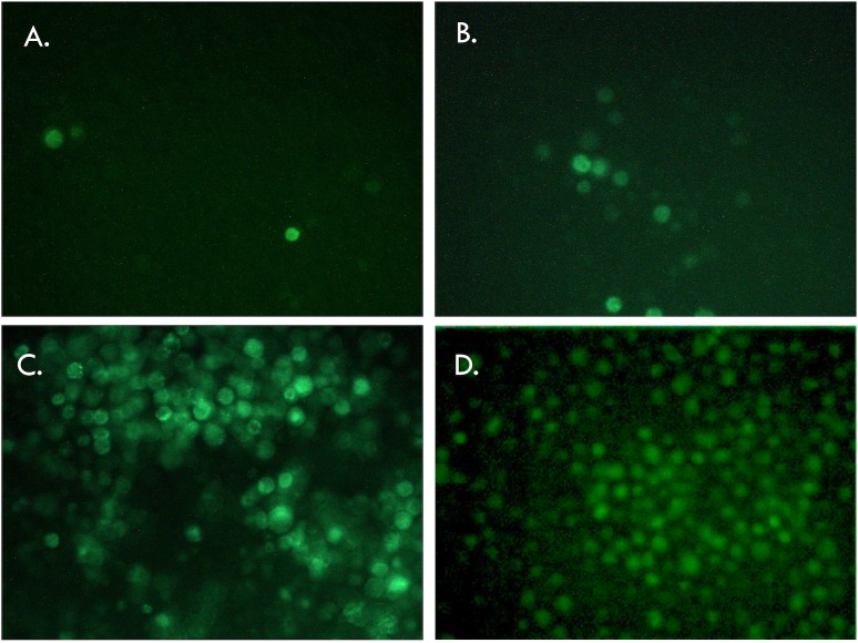
Fig 2. Fluorescence microscopic observation of Sf9 cells transfected with bacmid containing the Rep78eGFP cassette and expressing GFP.
A, B and C, 30, 55 and 120 hours post transfection of bacmid containing the Rep78eGFP fusion protein under the native p5 promoter. GFP is visible after 30 hours post transfection, and the combination of baculovirus replication followed by cell infection led to near 100% of the cells being GFP positive 120 hours post transfection. D. 120 hours post transfection with the bacmid containing the construct Rep78eGFP delta ITR.

