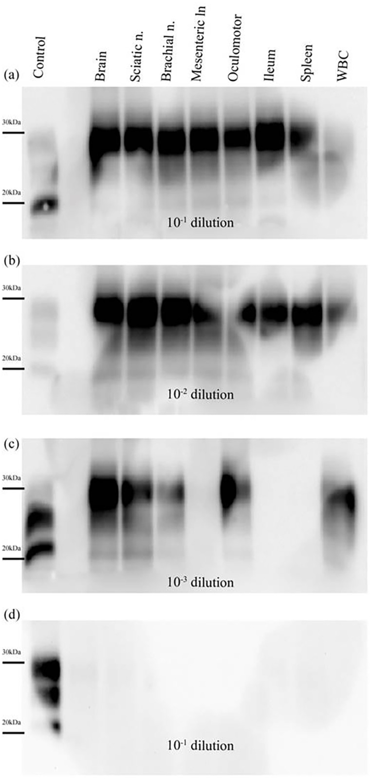
Fig 4. PrPres detection in PMCA reactions seeded with tissue samples collected from a Sh-BSE clinically affected pig.

Tissues: brain, sciatic nerve, brachial nerve, mesenteric lymph node, oculomotor muscle, ileum, spleen, and white blood cells (WBC). The animal was culled at 30 months after intracerebral inoculation with Sh-BSE. After 3 rounds of PMCA (48 hours each), PrPres amplification was detected at dilutions of 10−1 (a) and 10−2 (b) in all the tissues analysed. No PrPres was amplified in the mesenteric lymph node, ileum, or spleen at a dilution of 10−3 (c). Results obtained for 10−1 dilution after 3 rounds of PMCA in reactions seeded with tissues from a negative control pig (d). Control: PK digested classical scrapie isolate.
