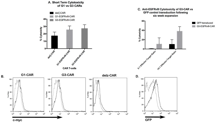
Fig 4. G1 vs G3 CAR and durability of cytoxicity in culture.
(A)Short term biophotonic cytolysis assay. Seven days after nucleofection of PBMCs with G1-EGFRvIII CAR (G1), G3-EGFRvIII CAR (G3), or delζ-EGFRvIII CAR [2] plasmids, cells were added to U87vIIIffluc cells at a 25:1 ratio. 5 hours after incubation, T-cell activity was measured by bioluminescence release, and percentage of specific release was calculated for each group. Mean cytolysis values ± SEM is shown for n = 3 for all groups. (B)Flow cytometric detection of G1-EGFRvIII, G3-EGFRvIII, or delζ-EGFRvIII CAR cell surface expression on PBMC transfectants 7 days post nucleofection. Expression of c-myc was typically detected as low magnitude population shift of mean fluorescence of 0.5 log units. (C)Post-culturing cytolysis in PBMCs that were nucleofected with G3-MR1 plasmids and cocultured with irradiated CD32-80-137L-EGFRVIIIΔ654 for 6 restimulation cycles (7-day cycles). Cells were added to U87vIIIfluc target cells at 5:1 or 10:1 ratios. Mean cytolysis values ± SEM is shown for n = 3 for all groups. (D)Flow cytometric detection of GFP in control PMG-GFP-T cells following six-week expansion. GFP expression was observed as a low fluorescence intensity shift of the entire transduced population versus control cells.

