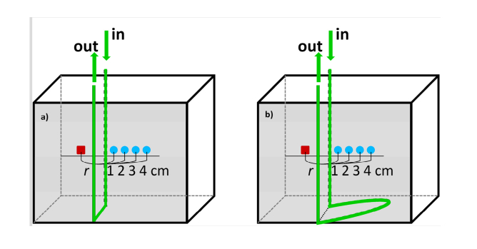
Fig. 3.
Geometry of the liquid phantom with a) short loop, b) long loop between segments of the tube located deeper and superficially. Boli with indocyanine green (ICG) were passing through the tube positioned in the fish tank at two depths in respect to the front wall of the phantom. Source fiber (red square) and detection fiber bundle (blue circle) were fixed on the surface of Mylar film which formed the front wall of the phantom. The segment of the tube located deeper was positioned at depth of 2 cm in respect to the front wall of the phantom.

