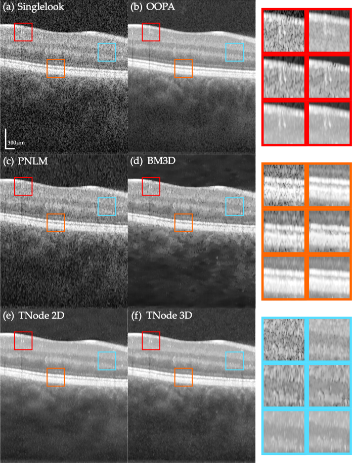
Fig. 3.
Native single-look cross-sectional view of human retina in vivo (a) and corresponding images despeckled with (b) OOPA, (c) PNLM, (d) BM3D, (e) TNode 2D and (f) TNode 3D. The insets at right are organized in the same order. See Visualization 1 (49.8MB, avi) for a comparison of the full tomogram.

