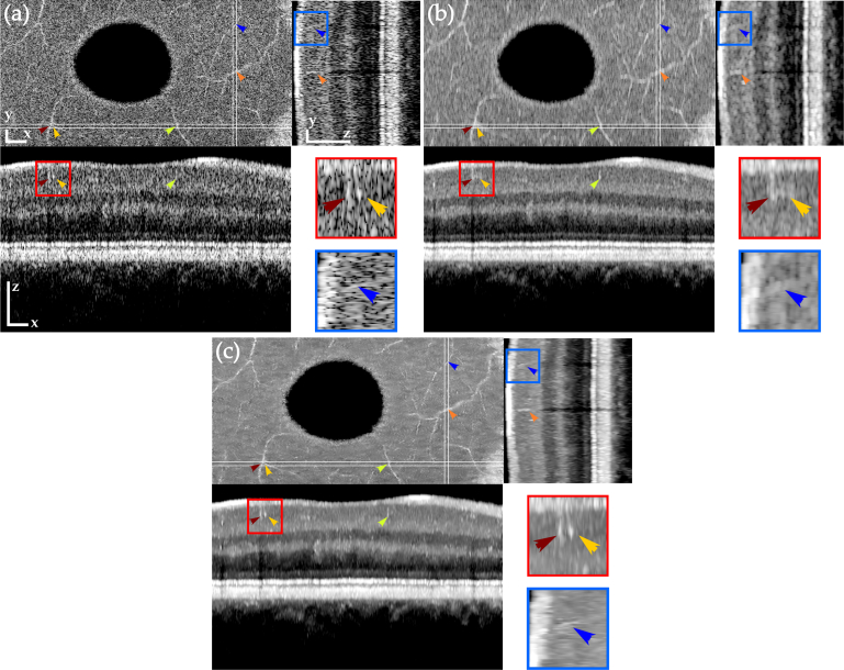
Fig. 4.
Orthogonal views of tomograms before (a) and after despeckling using OOPA (b) and TNode 3D (c). TNode preserves the two speckle-sized vessels shown in the red box as two independent structures, while OOPA merges them into a single structure. Resolution in the yz projection (small blood vessel in blue box) is mostly preserved in TNode. Scale bars correspond to 300μm in each direction.

