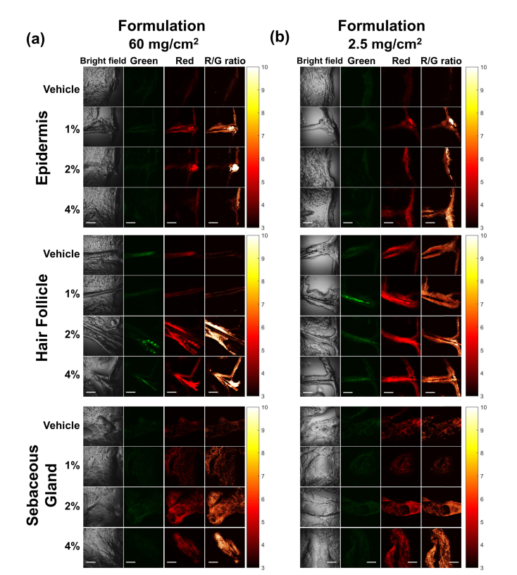
Fig. 2.
TPEF images of facial skin samples treated with (a) 60 mg/cm2 and (b) 2.5 mg/cm2 of BPX-01 formulation with 0% (vehicle), 1%, 2%, and 4% of MNC-Mg2+ for 24 hours at three anatomical sites: epidermis, hair follicle, and sebaceous gland. The TPEF images were acquired from two fluorescence channels: 445-480 nm (green) and 590-650 nm (red). The R/G ratio images were generated by dividing the red channel images by the green channel images on a pixel-by-pixel basis. The scale bar is 50 μm.

