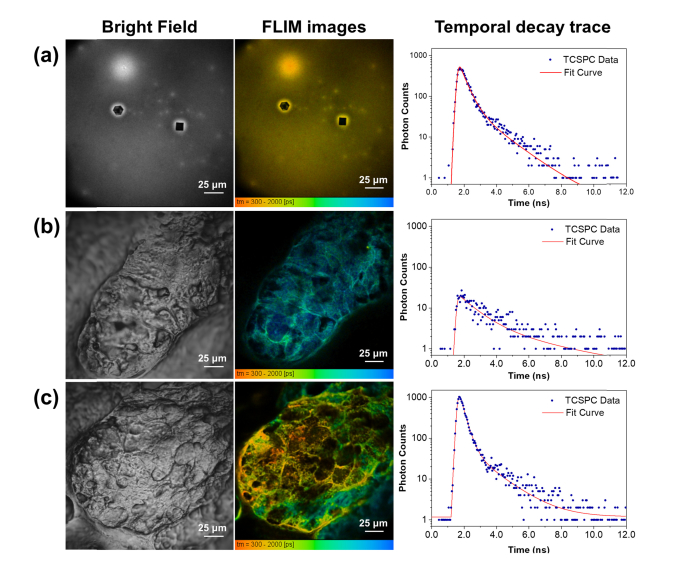
Fig. 3.
Bright field and FLIM images, as well as temporal decay traces of (a) dried MNC-Mg2+, (b) an untreated sebaceous gland, and (c) a sebaceous gland treated with MNC-Mg2+. The FLIM images were constructed from the red fluorescence channel (590-650 nm). All FLIM images were generated by fitting a double-exponential decay function to each pixel’s decay trace using SPCImage software.

