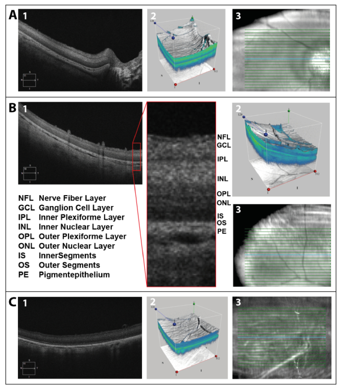
Fig. 1.
Central (A) and peripheral (B) OCT-scan approximately 1 h post mortem, as well as peripheral (C) scan 7 h post mortem: (1) retinal scan, (2) three-dimensional image of scanned area, and (3) location of depicted scan. The different retinal layers were labeled in the peripheral scan 1 h post mortem. The blue line in the right column shows the scan position shown in the left column.

