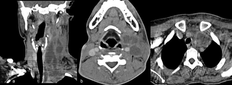
Fig. 2.

( A ) Contrast-enhanced computed tomography (CECT) neck soft tissue window coronal view showing multiple low attenuated central necrotic components with capsular ring enhancement and surrounding inflammatory changes suggestive of multiple abscess extending to the mediastinum. ( B ) Axial cut of a CECT of the neck showing a non-enhancing thrombosed internal jugular vein (arrow) with multiple abscesses and mass effect with effacement of the adjacent structures. ( C ) CECT of the mediastinum showing an abscess on the left side with mediastinal extension pushing the trachea to the right side.
