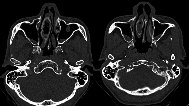
Fig. 1.

(a and b) Axial sections of computed tomography of the paranasal sinuses showing mucosal thickening within the right concha bullosa, and thickening of the surrounding bony walls. Also noted are areas of mild mucosal thickening in both maxillary sinuses and a mucosal polyp in the anterior wall of left maxillary sinus.
