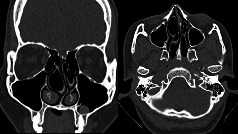
Fig. 3.

(a) Coronal and (b) axial sections of computed tomography of the paranasal sinuses show the presence of multiple air cells within the bilateral concha bullosae. Deviation of the nasal septum to the left side is noted. Also seen is a small mucosal polyp in the inferior wall of the left maxillary sinus.
