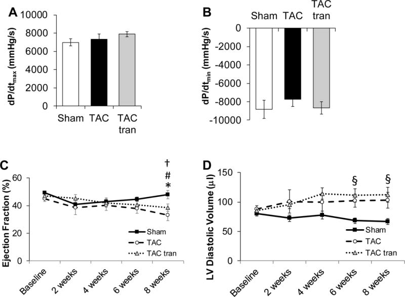
Figure 3. Assessment of cardiac function after 8 weeks of treatment with tranilast.

IVP measurements at 8 weeks post-TAC of maximum rate of contraction (dP/dtmax) (A) and maximum rate of relaxation (dP/dtmin) (B). Echocardiographic measurements of Ejection Fraction (C) and LV diastolic volume (D) at baseline, 2, 4, 6, and 8 weeks post-surgery (*P<0.05 TAC compared to sham; †P<0.05 TAC baseline vs TAC 8 weeks; #P<0.05 TAC tranilast baseline compared to TAC tranilast 8 weeks; §p<0.05 TAC tranilast compared to sham). P values are from one-way ANOVA (A and B) and 2-way ANOVA RM (C and D).
