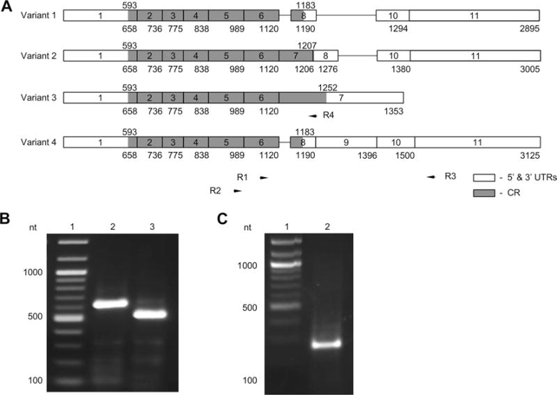
Fig. 1.

Rbpms splicing variants encoding isoforms A and C are expressed in human retina. a Splicing variants 1, 2, 3, and 4 are shown. Exons (boxes) are numbered from 1 to 11. Numbers under exons indicate the number of the last nucleotide of that exon. Positions of the coding regions (grey) are indicated above each splicing variant. Positions of primers (R1, R2, R3, and R4) used in PCR are indicated by arrows. b PCR products (lanes 2 and 3) corresponding to variant 1. PCR was performed with primer pairs R2/R3 (lane 2) and R1/R3 (lane 3). c PCR with primers R2/R4 produce a product corresponding to variant 3. Lane 1; DNA ladder
