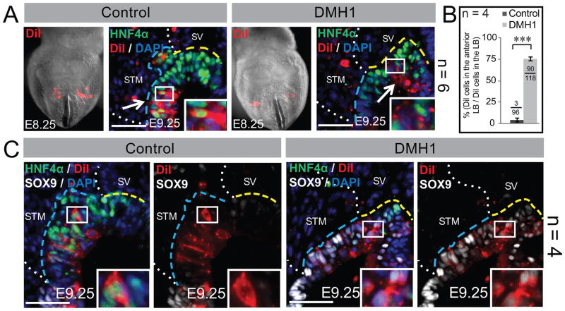
Figure 2. Anterior displacement of the putative posterior liver bud upon BMP inhibition.
A, C) Control or DMH1 treated embryos are DiI-labeled (red) and cultured. (A) Frontal views of whole embryos with DiI-labeled lateral hepatic progenitors at the onset of culture (E8.25). Section immunofluorescence after culture (E9.25) confirms that the lateral progenitors of control embryos mainly contribute to HNF4α+ hepatoblasts in the posterior liver bud. In contrast, the DiI-labeled lateral progenitors of DMH1 treated embryos are displaced towards the SV-bounded anterior liver bud and mainly do not express HNF4α. The arrows point to the bulk of the DiI-labeled cells and each inset is a higher magnification of the boxed area. (B) A quantification of the anteriorly displaced DiI-labeled cells as a percent of the total DiI-labeled cells in the whole liver bud. The fraction at the top of each bar depicts the number of the DiI-labeled cells counted in the anterior over the total number of DiI-labeled cells in the liver bud. (C) Section immunofluorescence of the DiI-labeled controls after culture, reveals that the DiI-labeled cells are not only restricted to the posterior liver bud but that these cells mainly express HNF4α and restrict the pancreatobiliary marker, SOX9. After DMH1 treatment, the anteriorly displaced DiI-labeled cells do not express HNF4α, but instead, aberrantly express SOX9. The insets reveal a higher magnification of the boxed area. The number of embryos (n) used for each experiment is listed to the right of each panel or within the graph. Each scale bar = 50 μM. Annotations are as in Figure 1.

