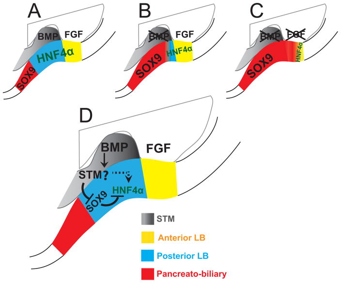
Figure 7. Working model for the role of BMP and FGF signals in hepatic induction.
A) Under the influence of normal BMP and FGF signaling the posterior foregut endoderm is divided into discrete domains: the anterior liver bud (yellow), in contact with the SV, the posterior liver bud (blue), in contact with the STM and the pancreatobiliary domain (red) which lies immediately posterior to the liver bud and mostly outside the STM. (B) Upon loss of BMP signals the hepato-pancreatobiliary boundary becomes diffuse; the posterior liver bud is lost and SOX9 is present in the posterior hepatic domain. (C) Upon loss of both BMP and FGF signaling almost all of the HNF4α+ liver bud is lost and ectopic SOX9 is observed throughout the hepatic domain. (D) Signaling events likely occurring in the hepatic domain and surrounding tissues. BMP promotes HNF4α in the posterior liver bud through indirect mechanism/s (possibly through the STM; the question mark indicates query). At least one indirect function of BMP is to repress SOX9 in the hepatic domain where SOX9 can repress HNF4α. Other unknown mechanisms may exist depicted by the dashed arrow. Annotations are as in Figure 1.

