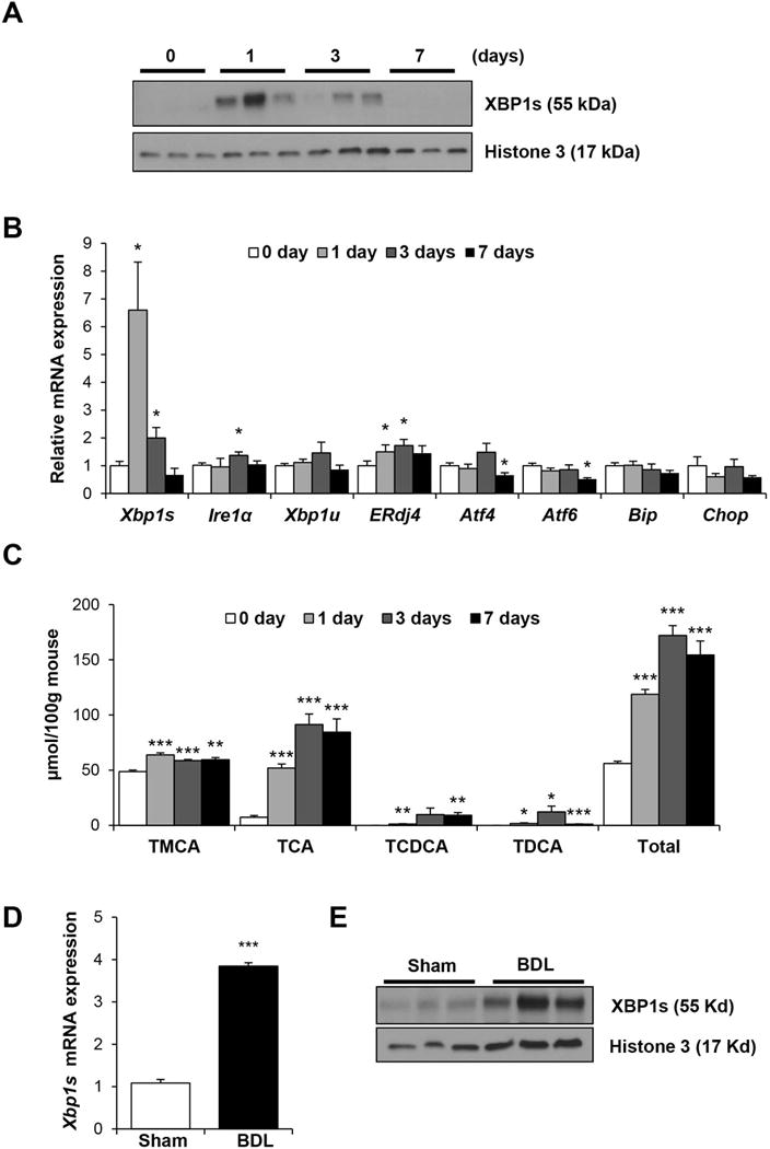
Figure 1. Hepatic UPR gene expression and bile acid pool in mice fed deoxycholic acid (DCA) and subjected to bile duct ligation (BDL).

Male FVB/NJ mice were fed chow supplemented with 0.3% DCA for 1, 3 and 7 days or chow-alone (0 day) (n=5). A) Western blot of hepatic nuclear XBP1s is shown with histone 3 used as a loading control. B) Hepatic gene expression was measured using qPCR. DCA-fed mice have increased hepatic gene expression of Xbp1s, ERdj4, and Ire1α. C) Bile acid composition and total bile acid pool in DCA- and chow-fed mice. D) Female C57BL/6J mice were subjected to BDL or sham operation (n=3) for 48 hours. Hepatic Xbp1s mRNA was measured by qPCR. E) Western blot of hepatic nuclear XBP1s is shown with histone 3 used as a loading control. *P < 0.05, **P < 0.01, ***P < 0.001 compared to 0 day or sham-control.
