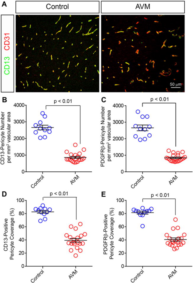
FIG. 2.

Vascular pericytes are reduced in human bAVMs. Representative confocal microscopy analysis (A) of CD13-positive pericytes (green) and CD31-positive endothelium (red) in human AVMs and temporal cortex from NVLCs. Graphs showing quantification of CD13-positive (B) and PDGFRβ-positive (C) pericyte cells per square millimeter vascular surface area in NVLCs and bAVMs. Graphs showing quantification of pericyte coverage of the vascular wall utilizing CD13 (D) and PDGFRβ (E) immunolabeling of pericyte cell processes. Values in the graphs are expressed as the mean ± standard error of the mean.
