
FIG. 5.
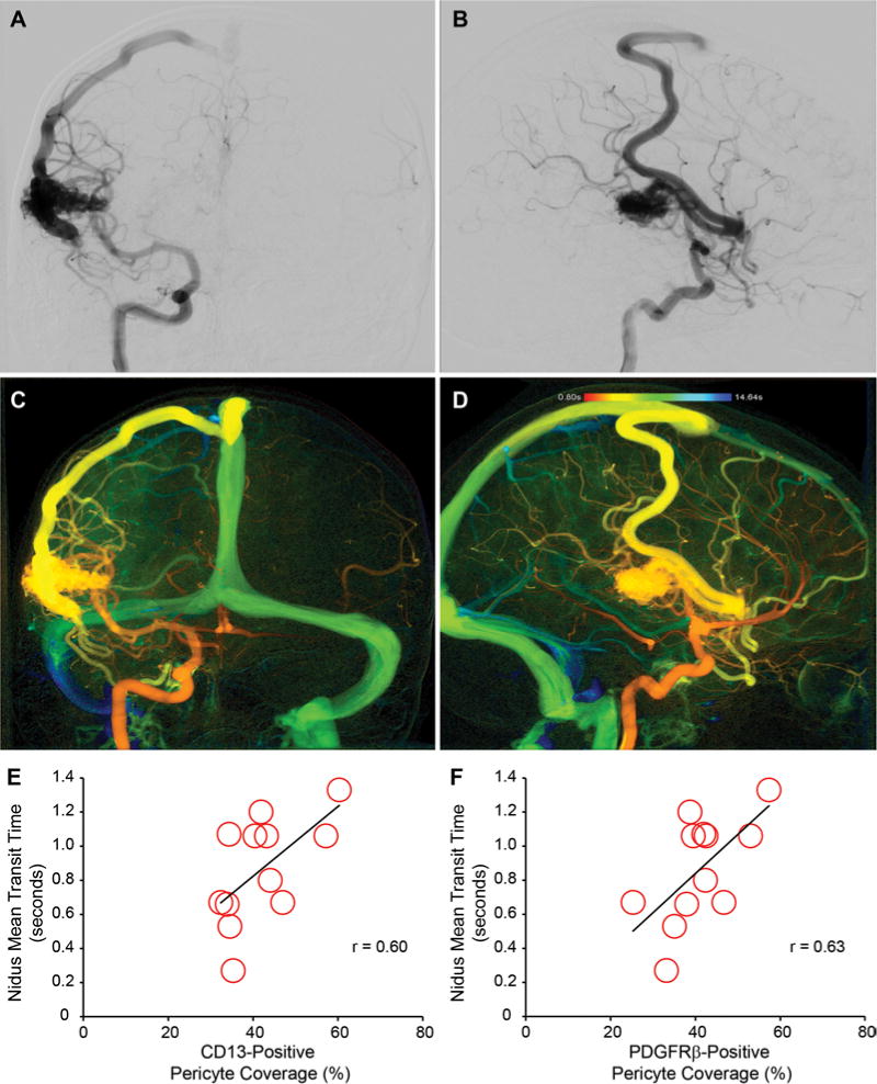
Pericyte reductions positively correlate with blood flow through the bAVM nidus. Representative preoperative angiograms, anteroposterior (A) and lateral (B) views of left internal carotid artery injection, demonstrating Spetzler-Martin grade II, supplementary grade 4 right lateral temporal bAVM. Representative syngo iFlow postprocessing of preintervention angiogram to determine mean iFlow transit time of bAVM nidus, anteroposterior (C) and lateral (D) views of left internal carotid artery injection. Graphic representations of correlation between CD13-positive (E) or PDGFRβ-positive (F) pericyte coverage and MTT of blood flow through the bAVM nidus using iFlow analysis. Each point depicts an individual subject.