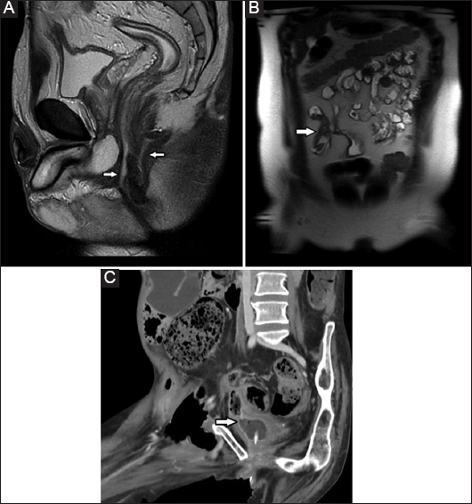
Figure 2.

(A) Sagittal T2-weighted single-shot fast spin-echoic image of the pelvis shows complex perianal sepsis with two fistulous tracts arising from the anterior and posterior aspect of the anal canal (arrows); (B) Coronal T2-weighted single-shot fast spin-echo image of the abdomen shows two adjacent small bowel loops tethered to each other, indicative of an enteroenteric fistula (arrow), known as the “star-sign”; (C) Coronal computed tomography enterographic section shows a fistula between an inflamed segment of small bowel and the urinary bladder (arrow)
