Fig. 2.
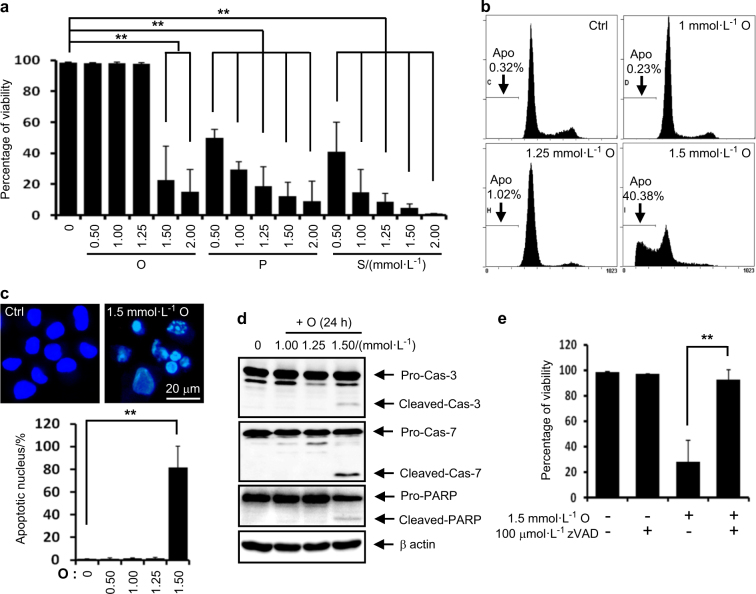
FFAs exert lipotoxicity in rat articular chondrocytes. a Viability assay. Palmitate (P) or stearate (S) at 0.5–2 mmol·L-1 and oleate (O) at 1.5–2.0 mmol·L-1 significantly reduced cell viability (n = 4). **P < 0.01 vs. vehicle according to Scheffe’s test. b Representative histograms showing cell cycle progression and the induction of apoptosis (Apo, the percentage of the population undergoing apoptosis). Oleate at 1.5 mmol·L-1 markedly increased the number apoptotic cells. c Representative Hoechst staining. The quantification of staining demonstrates that 1.5 mmol·L-1 oleate significantly increased the number of cells with condensed apoptotic nuclei. **P < 0.01 vs. vehicle according to Scheffe’s test. Scale bar, 20 μm. d Representative western blots showing that 1.5 mmol·L-1 oleate significantly induced the activation of apoptosis-related factors (n = 4). e Viability assay showing that zVAD-fmk significantly prevented 1.5 mmol·L-1 oleate-induced cell death (n = 4). **P < 0.01 according to Scheffe’s test

