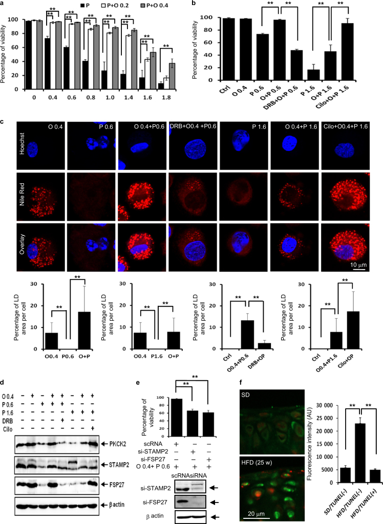
Fig. 7.
LD accumulation rescues articular chondrocytes co-incubated with palmitate and oleate in vitro and articular chondrocytes of mice fed an HFD in vivo. a Viability assay showing that 0.2 or 0.4 mmol·L-1 oleate supplementation suppressed palmitate-induced lipotoxicity (n = 4). **P < 0.01 vs. experimental control (palmitate alone treatment) according to Scheffe’s test. b Viability assay. Oleate at 0.4 mmol·L-1 significantly reversed 0.6 and 1.6 mmol·L-1 palmitate-induced lipotoxicity. PKCK2 inhibition by DRB and PKCK2 augmentation by cilostazol showed reciprocal influences on lipotoxicity (n = 4). **P < 0.01 vs. experimental control according to Scheffe’s test. c Representative confocal microscopy images. Oleate supplementation induced LD accumulation in articular chondrocytes treated with 0.6 or 1.6 mmol·L-1 palmitate. While DRB reduced LD accumulation in cells treated with 0.4 mmol·L-1 oleate plus 0.6 mmol·L-1 palmitate, cilostazol augmented LD accumulation in cells treated with 0.4 mmol·L-1 oleate plus 1.6 mmol·L-1 palmitate. LD accumulation was associated with resistance to the lipotoxicity in chondrocytes co-treated with oelate and palimate (n = 4). **P < 0.01 vs. control or experimental control according to Scheffe’s test. Scale bar, 10 μm. d Representative western blots showing that the increase in the LD accumulation in chondrocytes co-treated with 0.4 mmol·L-1 oleate and 0.6 mmol·L-1 palmitate or 0.4 mmol·L-1 oleate and 1.6 mmol·L-1 palmitate was correlated with the increase in the expression level of PKCK2, STAMP2 and FSP27 (n = 4). e Viability assay showing that siSTAMP2 and siFSP27 significantly decreased the viability of chondrocytes co-treated with 0.4 mmol·L-1 oleate and 0.6 mmol·L-1 palmitate (n = 4). **P < 0.01 vs. scRNA-administered experimental control according to Scheffe’s test. f Representative TUNEL and BODIPY double-labelling on cartilages obtained from OA model without surgery. The BODIPY fluorescence intensity in articular chondrocytes of mice fed an SD is lower compared to TUNEL-negative cells of mice fed an HFD. The quantification of BODIPY staining showed that the total LD volume was significantly increased in TUNEL-negative cells of mice fed an HFD (n = 4). **P < 0.01 vs. articular chondrocytes of mice fed an SD according to Scheffe’s test. The BODIPY fluorescence intensity in TUNEL-positive cells of mice fed HFD is lower compared to TUNEL-negative cells. The quantification of BODIPY staining showed that the total LD volume was significantly reduced in TUNEL-positive cells (n = 4). **P < 0.01 vs. TUNEL-negative cells according to Scheffe’s test. Scale bar, 20 μm

