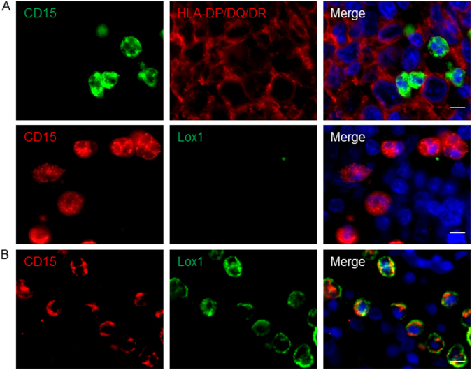
Fig. 2. Tumor-associated neutrophils from DLBCL patients display a mature blood neutrophil phenotype.
a In situ fluorescence co-staining of DLBCL biopsies for CD15 and HLA-DR (upper panel) or Lox1 (lower panel). b In situ fluorescence co-staining of tonsil for Lox1 and CD15. Hoechst33342 nuclear staining (blue) is also shown in the merge pictures. Pictures are representative of 10 DLBCL patients and three healthy tonsils. Scale bar = 10 µm

