Figure 3.
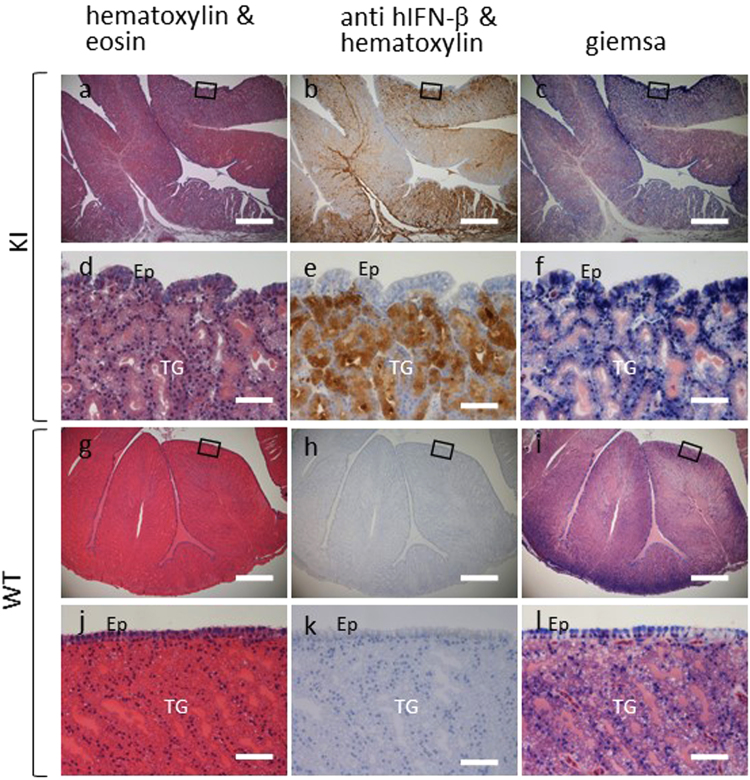
Immunohistochemistry of hIFN-β in the oviduct magnum sections. Oviduct sections from hIFN-β KI (a–f) and WT (g–l) hens were stained with hematoxylin and eosin (a,d,g and j), immunohistochemically stained for hIFN-β and counterstained with hematoxylin (b,e,h and k), or stained with Giemsa (c,f,i and l). Panels d–f and j–l are magnified views of the enclosed rectangular sections in panels a–c and g–i, respectively. The presence of hIFN-β is apparent in the oviduct magnum section from the hIFN-β KI hen (b,e) with its expression restricted to the tubular glands (TG). Ep, epithelial cells. All sections were counterstained with hematoxylin. Scale bars, 500 μm (a–c,g–i); 50 μm (d–f,j–l).

