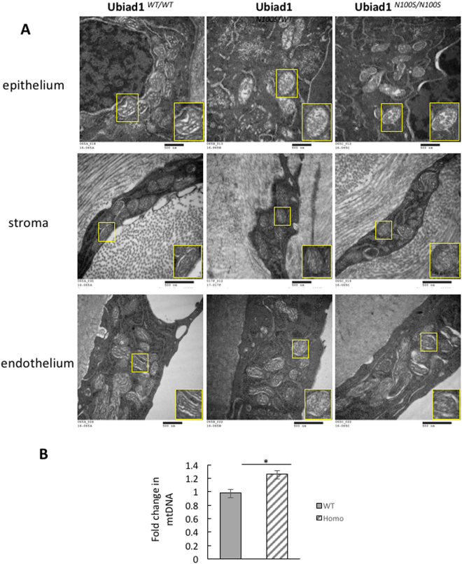
Figure 3.
Mitochondria damage in Ubiad1N100S cornea. (A) Electron Microscopy showed mitochondrial degeneration in the corneal epithelial cells, stroma keratocytes, and endothelium of heterozygous and homozygous mice. Inset at lower right in each micrograph shows an individual mitochondrion at higher magnification. Mice were 4-month-old males. (B) Quantification of mitochondrial DNA copy number change in wild type mice and Ubiad1N100S/N100S mice by real-time PCR. The fold change was 0.98 in wild type mice versus 1.27 in Ubiad1N100S/N100S mice, P < 0.05. Mice were of mixed genders.

