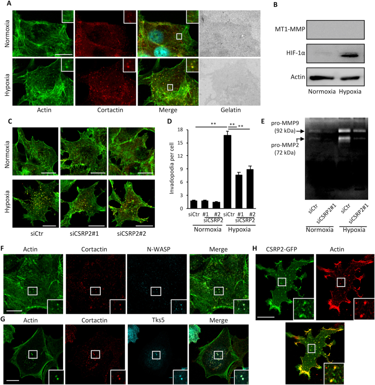
Figure 4.
Hypoxia-induced invadopodium precursors formation in MCF-7 cells requires CSRP2 upregulation. (A) Gelatin degradation assay showing that normoxic (upper panels) and hypoxic (lower panels) MCF-7 cells do not promote (Oregon Green 488-labelled) gelatin degradation after 48 h. After fixation, MCF-7 cells were stained for actin (in green) and cortactin (in red), and imaged using confocal microscopy. (B) Western blot analysis of the MT1-MMP and HIF-1α protein levels in normoxic and hypoxic MCF-7 cells. (C) MCF-7 cells were transfected with non-targeting or 2 different CSRP2 siRNAs (siCSRP2#1 and 2), cultured under normoxic or hypoxic conditions, and invadopodium precursors were detected by actin (green) and cortactin (red) co-labelling. (D) Quantitative analysis of invadopodium precursor density in MCF-7 cells as in (C). The data originate from 3 independent experiments (n = 60). (E) Gelatin zymography assay conducted with the conditioned media collected from MCF-7 cells cultured as in (C,D). The zymogram is representative of three independent experiments. Similar data were obtained with siCSRP2#2. (F,G) Hypoxic MCF-7 cells were labelled for actin (green), cortactin (red) and either N-WASP (immunolabelling, F) or Tks5 (Tks5-GFP, G). (H) CSRP2-GFP co-localizes with actin (red) at invadopodium precursors in hypoxic MCF-7 cells. The insets show magnification of invadopodium precursors. Bars = 15 µm; *p < 0.05; **p < 0.01.

