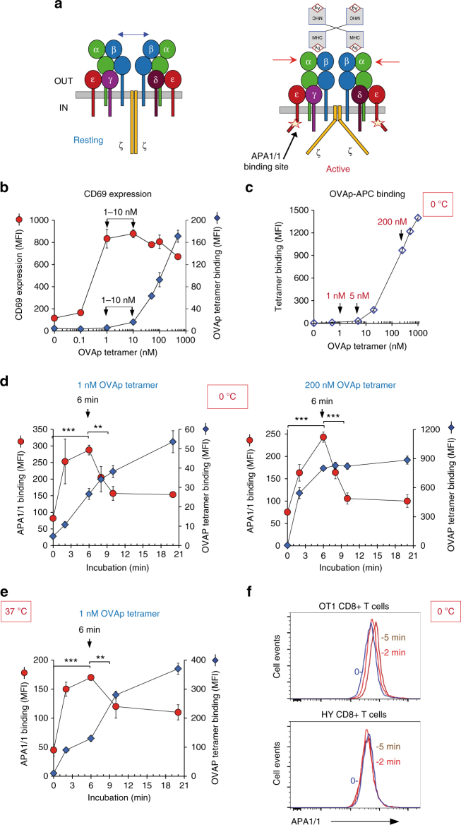
Fig. 1.
Optimum time for Active conformation of the TCR at sub-saturating concentrations of the pMHC ligand. a Cartoon of the Resting and ligand-bound Active conformations of the TCR. Bivalent or multivalent ligation of two or more TCRs by its pMHC ligand results in an outside-in transmission of a conformational change detected in the cytoplasmic tails of the CD3 subunits. One of these changes consists in exposing an epitope in the proline-rich region of CD3ε for mAb APA1/1 binding. b OT-1 CD8 + T cells were incubated at 37 °C with the indicated concentrations of APC-labelled OVAp tetramer for 24 h. After stimulation, cells were stained with anti-CD69 and analyzed by cytometry. The mean fluorescence intensity (MFI) for CD69 staining is shown as red circles and that of tetramer binding as blue diamonds. c OT-1 CD8 + T cells were incubated on ice with the indicated concentrations of APC-labelled OVAp tetramer for 40 min. MFI was calculated by flow cytometry. d OT-1 CD8 + T cells were incubated at different times at 0 °C with 1 nM or 200 nM of OVAp-APC tetramer and subsequently fixed, permeabilized and stained with the APA1/1 mAb prior to flow cytometry analysis. MFI for APA1/1 staining is shown as red circles and that of tetramer binding as blue diamonds. e OT-1 CD8 + T cells were incubated at different times at 37 °C with 1 nM OVAp-APC tetramer and stained with the APA1/1 mAb as in Fig. 1d. f Histogram overlay for APA1/1 expression in OT-1 and HY CD8 + T cells not incubated (blue lines) or incubated for 2 min (red line) or 5 min (brown line) with 1 nM OVAp-APC tetramer at 0 °C. All data shown in Fig. 1 represent the mean ± s.d. of triplicate datasets; *p < 0.05; **p < 0.005; ***p < 0.0005 (two-tail unpaired t-test)

