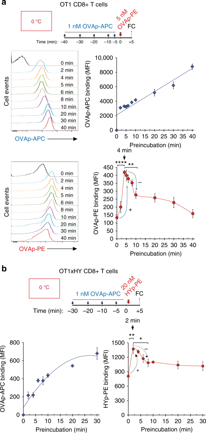
Fig. 3.

Binding of a first pMHC tetramer ligand at sub-saturating concentrations transiently favours binding of a second pMHC tetramer ligand. a OT-1 T CD8 + T cells were preincubated at 0 °C with 1 nM OVAp-APC tetramer for the indicated times and subsequently incubated with 5 nM of OVAp-PE tetramer for five additional minutes. Time 0 indicates binding of OVAp-PE tetramer in the absence of preincubation with OVAp-APC tetramer. Cells were fixed and stained for CD8 before analysis by flow cytometry and calculation of the MFI for OVAp-PE and OVAp-APC. Histograms for OVAp-APC tetramers and OVAp-PE tetramer fluorescence intensity are shown on the left panels. The MFI values for OVAp-APC tetramers and OVAp-PE tetramer are represented on the right panels. b CD8 + T cells from double transgenic female OT-1xHY mice were preincubated on ice with 1 nM OVAp-APC tetramer for the indicated times and subsequently incubated with 20 nM of HYp-PE tetramer for five additional minutes. Cells were fixed and stained for CD8 before analysis by flow cytometry to generate MFI values for HYp-PE and OVAp-APC binding. All data shown in Fig. 3 represent the mean ± s.d. of triplicate datasets; *p < 0.05; **p < 0.005; ****p < 0.00005 (2-tail unpaired t-test)
