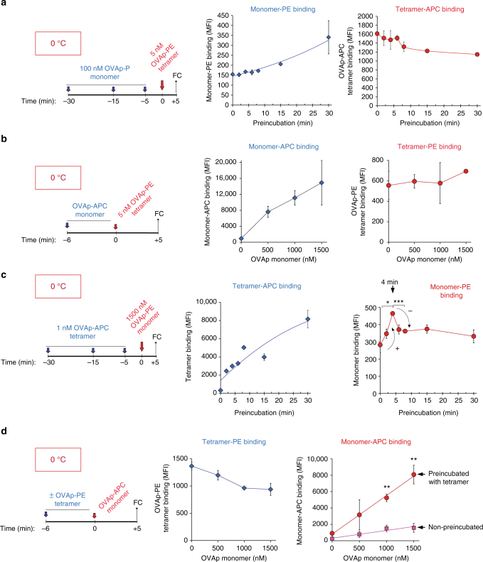
Fig. 4.
A multimeric pMHC ligand is required to promote cooperative pMHC binding. a CD8 + OT-1 T cells were preincubated at 0 °C with 100 nM biotinylated OVAp monomer for the indicated times and subsequently incubated with 5 nM of OVAp-APC tetramer for five additional minutes. Time 0 indicates binding of OVAp-APC tetramer in the absence of preincubation with OVAp monomer. Cells were fixed and stained with streptavidin-PE in order to detect binding of the monomer. Plots represent the MFI values for OVAp-PE monomer and OVAp-APC tetramer binding. b CD8 + OT-1 T cells were incubated at 0 °C with the indicated concentrations of biotinylated OVAp monomer for 6 min and subsequently incubated with 5 nM of OVAp-PE tetramer for 5 additional minutes. Cells were then fixed and stained with streptavidin-APC in order to detect the monomer. Plots represent the MFI values for OVAp-APC monomer and OVAp-PE tetramer binding. c CD8 + OT-1 T cells were preincubated at 0 °C with 1 nM OVAp-APC tetramer for the indicated times and subsequently incubated with 1.5 μM of biotinylated OVAp monomer (detected with streptavidin-PE) for 5 additional minutes. Time 0 indicates binding of OVAp monomer in the absence of preincubation with OVAp-APC tetramer. Cells were fixed and stained with streptavidin-PE in order to detect binding of the monomer. Plots represent the MFI values for OVAp-PE monomer and OVAp-APC tetramer binding. d CD8 + OT-1 T cells were preincubated or not at 0 °C with 1 nM of OVAp-PE tetramer for 6 min prior to incubation with the indicated concentrations of biotinylated OVAp monomer for five additional minutes. Cells were fixed and stained with streptavidin-APC in order to detect binding of the monomer. Plots represent the MFI values for OVAp-APC monomer and OVAp-PE tetramer binding. All data shown in Fig. 4 represent the mean ± s.d. of triplicate datasets; * p < 0.05; **p < 0.005; ***p < 0.0005 (2-tail unpaired t-test)

