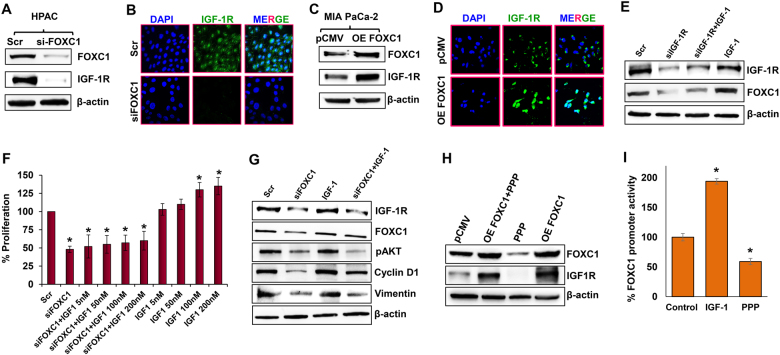
Fig. 2. Crosstalk between FOXC1 and IGF-1R in pancreatic cancer cells.
a Western blot analysis of FOXC1 and IGF-1R expression in FOXC1-silenced HPAC cells. b Immunofluorescence analysis of IGF-1R expression in FOXC1-silenced HPAC cells visualized through confocal microscopy at × 60 magnification. c Western blot analysis of FOXC1 and IGF-1R expression in FOXC1-overexpressing MIA PaCa-2 cells. d Immunofluorescence analysis showed increased IGF-1R expression in FOXC1-overexpressing MIA PaCa-2 cells (× 60 magnification). e Western blot analysis of FOXC1 and IGF-1R expression in HPAC cells treated with scramble siRNA (Scr) or IGF-1R siRNA or IGF-1R siRNA plus 100 nM of IGF-1 or 100 nM of IGF-1 alone. f MTS assay was used to measure the cell proliferation in response to IGF-1 treatment (5, 50, 100, and 200 nM) in parental and FOXC1-silenced HPAC cells. g Western blot analysis of IGF-1R, FOXC1, pAKT, Cyclin D1, and Vimentin in HPAC cells treated with FOXC1 siRNA or IGF-1 or FOXC1 siRNA plus IGF-1. h Western blot analysis of FOXC1 and IGF-1R expression in parental and FOXC1-overexpressing MIA PaCa-2 cells treated with or without Picropodophyllin (PPP). i Gaussia luciferase assay was used to show FOXC1 promoter activity in MIA PaCa-2 cells in the presence of IGF-1 or PPP. Data shown as mean ± SEM. Experiments (n = 3) were repeated three times in triplicates. *p < 0.05

