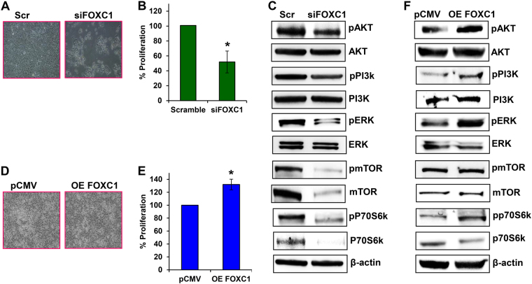
Fig. 3. Effect of FOXC1 on pancreatic cancer cell proliferation.
a Morphological changes occurred in FOXC1-silenced HPAC cells, which were observed under a light microscope at × 10 magnification. b MTS assay was used to measure cell proliferation of FOXC1-silenced HPAC cells. c Western blot analysis of proliferation markers in FOXC1-silenced HPAC cells. d Morphological changes occurred in FOXC1-overexpressing MIA PaCa-2 cells, which were observed under a light microscope at × 10 magnification. e MTS assay was used to measure the proliferation of FOXC1-overexpressing MIA PaCa-2 cells. f Western blot analysis of proliferation markers in FOXC1-overexpressing MIA PaCa-2 cells. Results represented as mean ± SEM. Data shown as mean ± SEM. Experiments (n = 3) were repeated three times in triplicates. *p < 0.05

