Figure 1.
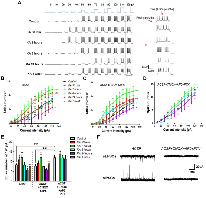
The neuronal excitability changed after kainic acid (KA) injection. (A) On the cortex of mice brain slices, depolarization currents with various intensities were injected into individual neurons to induce excitatory effect. Representative traces of the resting membrane potential (RP) baseline and evoked action potentials (spikes). (B) Evoked action potentials were quantified to compare the neuronal excitability at different time points after KA injection (n = 10–13 cells from 3–4 mice in each group). (C) Action potentials were evoked by depolarization currents and quantified in the presence of 20 μM CNQX and 50 μM AP5 (n = 11–13 cells from three mice in each group). (D) Action potentials evoked and quantified in the presence of 20 μM CNQX, 50 μM AP5 and 100 μM PTX (n = 9–13 cells from two mice in each group). (E) The number of spikes stimulated by 120 pA depolarization current was quantified in different conditions. *P < 0.05, **P < 0.01, ***P < 0.001 compared to control in the same recording condition. ##P < 0.01 of control mice compared between different conditions. (F) Spontaneous excitatory (sEPSCs) and inhibitory (sIPSCs) postsynaptic currents were recorded in the absence or presence of CNQX, AP5 and PTX.

