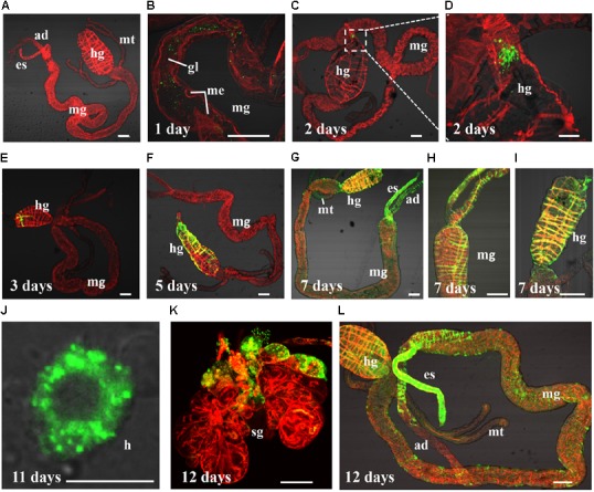
FIGURE 1.

Barley yellow striate mosaic virus (BYSMV) infection starts in the hindgut of SBPHs. Internal organs of infected SBPHs were immunolabeled for BYSMV with N-FITC (green) and stained for actin with phalloidin-rhodamine (red), then examined by confocal microscopy. (A) View of dissected alimentary canal of a negative control planthopper unexposed to BYSMV. At 1 (B), 2 (C,D), 3 (E), 5 (F), 7 (G–I), 11 (J), and 12 (K,L) days padp, internal organs of BYSMV-infected SBPHs were dissected, isolated, and processed for iCLSM. These images are representative of multiple experiments with multiple preparations. ad, anterior diverticulum; mg, midgut; hg, hindgut; mt, malpighian tubules; es, esophagus; sg, salivary gland; h, hemocytes; gl, gut lumen; me, midgut epithelium. Bar in hemocytes is equal to 10 μm. Other Bars, 100 μm.
