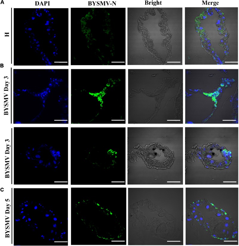
FIGURE 2.

Barley yellow striate mosaic virus accumulations in the cytoplasm of the hindguts of viruliferous planthopper vectors. The paraffin sections of the infected hindguts were immunolabeled for BYSMV with N antigens-specific IgG conjugated directly to fluorescein-5-isothiocyanate (green) and stained for nuclei with DAPI (blue). (A) The hindgut sections exposed to healthy plants served as negative controls. (B) At 3-day padp, BYSMV accumulations in the cytoplasm of epithelial cells of the hindguts of viruliferous planthopper vectors. Upper panel was vertical section; bottom panel was cross section. (C) At 5-day padp, BYSMV accumulations in the visceral muscles surrounding the infected hindguts. Bars, 50 μm.
