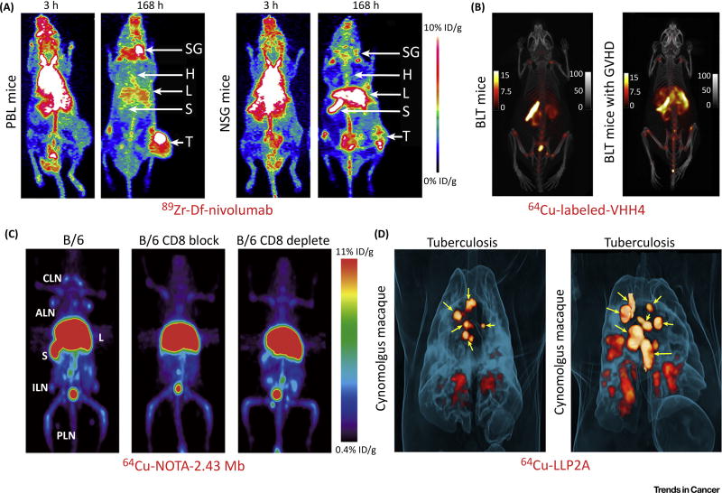
Figure 2. Positron Emission Tomography (PET) Imaging with Labeled Antibodies, Antibody Fragments, or Proteins Detects T Cells.
(A) 89Zr-Df-nivolumab successfully maps the in vivo biodistribution of tumor-infiltrating T cells expressing PD-1 in tumor-bearing mice. Note that tumor uptake of the tracer was much higher in humanized PBL tumor-bearing mice (left) than that in NSG tumor-bearing mice (right), which indicated infiltration of tumor-infiltrating lymphocytes into the tumor microenvironment in PBL mice. (B) 64Cu-labeled VHH4 PET imaging of BLT mice (left) and BLT mice with stage 3 graft-versus-host disease (GVHD) (right). The results showed that BLT mice with GVHD had intense PET signal in the liver, which indicated T cell infiltration in the affected liver. (C) ImmunoPET imaging of T cells using 64Cu-NOTA-2.43 minibody (Mb) in antigen-blocked and antigen-depleted B/6 mice. (D) 64Cu-LLP2A PET imaging of cynomolgus macaques infected with the Mycobacterium tuberculosis strain Erdman. Maximum intensity projections showed that 64Cu-LLP2A uptake in granulomas and infected lymph nodes before necropsy. Yellow arrows indicate lymph nodes. Adapted, with permission, from [28,37,57,60]. Abbreviations: ALN, axillary lymph nodes; B, bone; B/6, C57BL/6; CLN, cervical lymph nodes; H, heart; ILN, inguinal lymph nodes; L, liver; PLN, popliteal lymph nodes; S, spleen; SG, salivary gland; T, A549 tumor.

