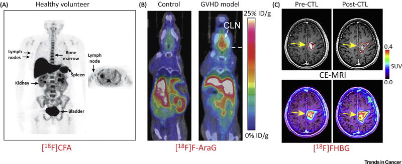
Figure 3. Molecular Imaging of T Cells via Radiolabeled Small Molecules or Positron Emission Tomography (PET) Reporter Probes.
(A) A first-in-human study of [18F]CFA, a nucleoside analogue probe, showed significant probe accumulation in deoxycytidine kinase (dCK)-positive tissues (bone marrow, liver, and spleen) and in the axillary lymph nodes of healthy volunteers, indicating its potential to delineate T cells. (B) [18F]F-AraG is an analog of AraG, a compound identified to have specific cytotoxicity toward T lymphocyte and T lymphoblastoid cells versus other immune cell types. [18F]F-AraG PET showed visibly higher tracer uptake in the cervical lymph nodes (CLN) in graft-versus-host disease (GVHD) models. (C) Contrast-enhanced MRI (CE-MRI) and [18F]FHBG PET imaging of recurrent glioma. T1-weighted CE-MRI demonstrated that the tumor size shrank after cytotoxic T cell (CTL) injections (top left panel). Accordingly, the [18F]FHBG signal was significantly increased after CTL infusions, with a 66% increase in total [18F]FHBG activity. Adapted, with permission, from [91,96,109]. Abbreviation: SUV, standardized uptake value.

