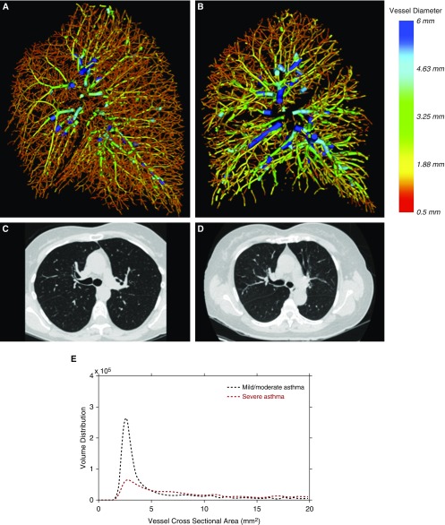
Figure 1.
Sagittal view of the three-dimensional quantitative reconstruction of the pulmonary vasculature in the right lung of (A) a subject without evidence of pruning and normal lung function (FEV1 percent predicted, 97%), and (B) a subject with evidence of pruning and impaired lung function (FEV1 percent predicted, 75%). The color red corresponds to smaller vessels, and blue corresponds to larger vessels. (C and D) Representative axial computed tomography images from the same two participants. (E) Quantitative profiles of blood volume distribution of the same two participants.

