-
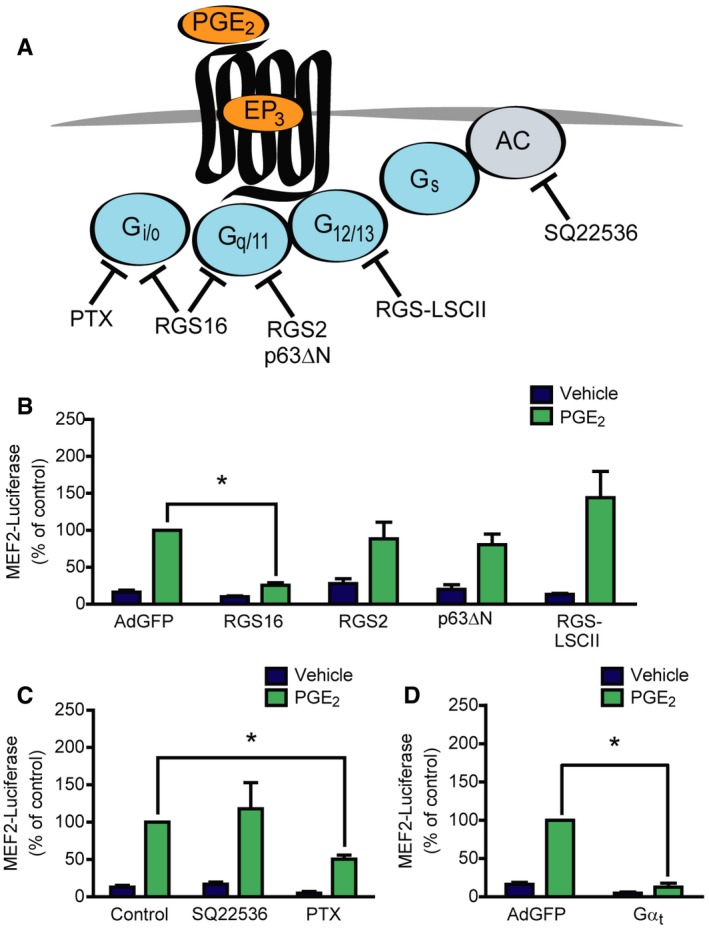
A
Illustration of the target points of the used pharmacological and protein‐based inhibitors.
-
B–D
(B, D) NRVMs were infected with the 3xMEF2‐Luciferase reporter and with recombinant adenoviruses encoding EGFP (AdGFP), RGS16, RGS2, p63ΔN, RGS‐LSCII, or Gαt, as indicated. The cells were serum starved for 20 h and stimulated with DMSO or 1 μM PGE2 for 24 h. (C) NRVMs were infected with the 3xMEF2‐Luciferase reporter and pretreated with the adenylyl cyclase inhibitor SQ22536 (100 μM) for 20 min or the Gi/o protein inhibitor pertussis toxin (PTX, 100 ng/ml) for 20 h in serum‐starved conditions and treated with DMSO or 1 μM PGE2 for 24 h.
Data information: In (B–D),
n =
3, independent experiments, *represents significant interaction between the two treatments (
P <
0.05, two‐way ANOVA, RGS16,
P <
0.0001; RGS2,
P =
0.1725; p63ΔN,
P =
0.0666; RGS‐LSCII,
P =
0.0818, SQ22536,
P =
0.385; PTX,
P <
0.0001; Gα
t,
P <
0.0001), values are mean ± s.e.m.
Source data are available online for this figure.