-
A
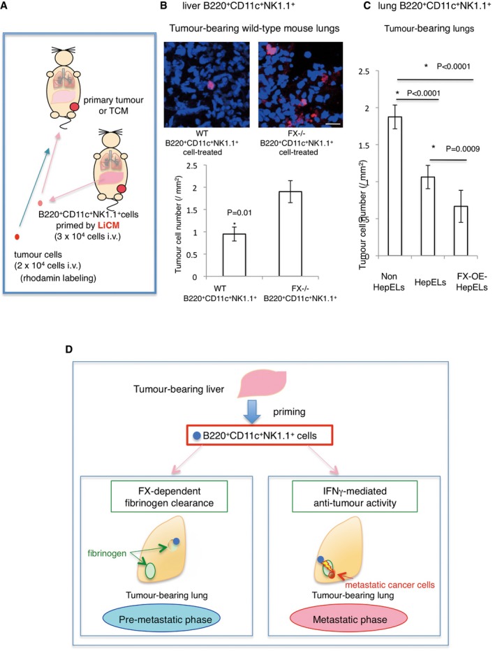
The experimental scheme. B220+CD11c+NK1.1+ cells derived from tumour‐bearing mouse liver or lungs were primed with LiCM or tethered with recombinant FX and then injected into tumour‐bearing or TCM‐stimulated mice; 24 h after the injection, tumour cells were injected. Tissues were analysed 48 h after the tumour cell injection.
-
B
Tumour cell homing in the lungs pre‐treated with LiCM‐primed B220+CD11c+NK1.1+ cells from tumour‐bearing WT or FX−/− mouse liver. Representative photographs of homing of rhodamine‐labelled tumour cells (upper, scale bar, 20 μm). Number of homing tumour cells are shown (lower). Shown are averages (N = 60 sections, 5/group, all field count/section, 12 sections/sample) with SEM and Welch's t‐test.
-
C
Tumour cell homing in the lungs pre‐treated with activated FX‐overexpressed B220+CD11c+NK1.1+ cells (FX‐OE‐HepELs) from tumour‐bearing lungs. Number of homing tumour cells after an injection of lung HepELs or FX‐OE‐HepELs is shown. Shown here are averages (N = 24 sections, 4/group, all field count/section, 6 sections/sample) SEM and one‐way ANOVA.
-
D
Model of B220+CD11c+NK1.1+HepEL functions in the pre‐ and post‐ metastatic phases. In the pre‐metastatic phase, tumour‐stimulating liver induces FX expression in B220+CD11c+NK1.1+ cells; then, the cells move into lungs to eliminate focal fibrinogens (left panel). In the post‐metastatic phase, if circulating tumour cells reach the fibrinogen area, B220+CD11c+NK1.1+HepELs attack tumour cells with IFNγ (right).

