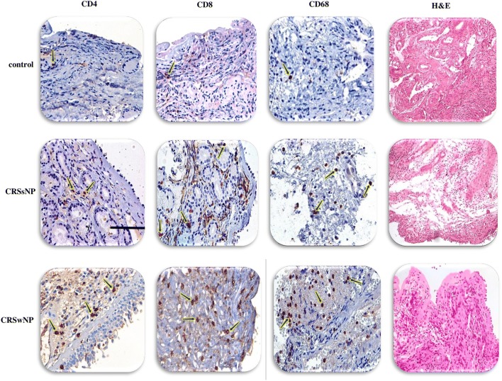
Fig. 1.
The distribution of infiltrating inflammatory cells to the sinonasal tissues. Representative IHC staining of CD4+ T cells, CD8+ T cells, and CD68+ macrophages, as well as H&E staining for eosinophils, neutrophils, and total inflammatory cells in controls, CRSsNP, and CRSwNP patients. Scale bar 100 µm. CRSsNP chronic rhinosinusitis without polyp, CRSwNP chronic rhinosinusitis with polyp

