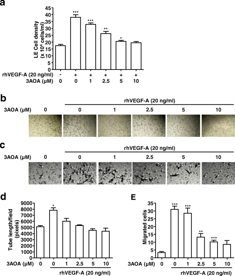
Fig. 3.

Effects of 3AOA on proliferation, tube formation, and migration in HLMECs stimulated with rhVEGF-A. a, Proliferation in HLMECs stimulated rhVEGF-A. Cells were detached and counted using a hemocytometer. b and d, Tube formation in HLMECs stimulated rhVEGF-A. Cells were imaged under a inverted phase contrast microscope using a digital single-lens reflex camera. Total tube lengths of a unit area were calculated using the Image J program. c and e, The migrated cells to the underside of membranes were fixed with methanol, stained with hematoxylin solution, and then imaged under a inverted phase contrast microscope using a digital camera. Five digital images per well for (C) were obtained, and the numbers of migrated HLMECs were counted. Each sample was assayed in duplicate. Numbers of migrated HLMECs present in 320 mm2 are presented as a bar diagram. Data are presented as a mean ± S.D. of three independent experiments (*p < 0.05, **p < 0.01, ***p < 0.001)
