
Fig. 5. Requirement of AnxA1 and its receptor FPR2/ALX for MV-induced chondroprotection.
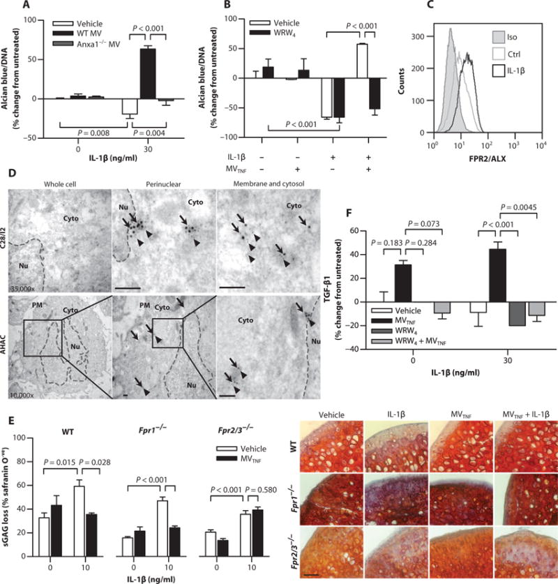
(A) ECM deposition normalized to DNA content in C28/I2 micromass cultures stimulated with IL-1β and cocultured with WT or Anxa1−/−mouse MVTNF (105). (B) ECM deposition normalized to DNA content in C28/I2 micromass cultures stimulated with a combination of IL-1β (30 ng/ml), human neutrophil-derived MVTNF (105), and WRW4 (10 μM, 10 min before MV addition). Data in (A) and (B) are means ± SEM (n = 4 separate MV and cell culture preparations). (C) FPR2/ALX expression in C28/I2 micromasses [stimulated as in (A)]. Data are representative of n = 3. (D) Immunogold labeling of AnxA1 (arrows) and FPR2/ALX (arrowheads) in C28/I2 and AHAC micromasses. Black boxes indicate panel magnified to the right for AHAC. Dashed lines, nuclear envelope. PM, plasma membrane; Cyto, cytoplasm; Nu, nucleus. Scale bars, 100 nm. (E) Cartilage explants from WT, Fpr1−/−, or Fpr2/3−/− mice were stimulated with IL-1β with or without 105 MVTNFs every other day for 7 days. Sections were stained with safranin O. Representative images of micrographs used for analysis are shown. Scale bars, 200 μm. Data are means ± SEM (n = 3 to 6). (F) Total TGF-β1 content in supernatants of C28/I2 micromasses treated as in (B). Data are means ± SEM (n = 8 separate MV donors). P values were determined by two-way ANOVA and Bonferroni posttest (A, B, E, and F).