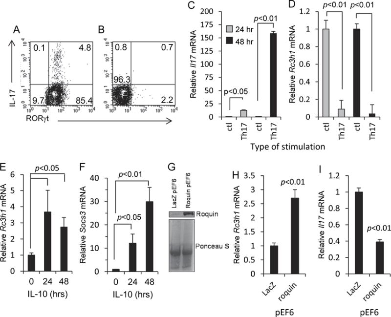
Fig. 1.

Opposing relationship between Roquin-1 and IL-17. (A) MLN T cells 48 hr after stimulation using the Th17 induction regimen described in the Materials and Methods. (B) MLN T cells 48 hr after CD3 stimulation in the absence of Th17 induction. Cells were gated onto the CD4+ cell population. Representative histograms from three experiments. (C, D) Increased Il17a and decreased Rc3h1 transcription in MLN T cells following 24 and 48 hr of Th17 induction. Control cultures consisted of plate-bound anti-hamster antibody with normal hamster IgG. (E) Increased Rc3h1 transcription in EL4 cells following exposure to IL-10. (F) Exposure of EL4 cells to IL-10 resulted in increased Socs3 transcription. (G) Western blot showing increased levels of Roquin-1 protein expression in EL4 cells transfected with the Roquin-1 pEF6 expression plasmid. Ponceau S staining was done to compare sample protein levels. (H, I) Roquin-1 pEF6 transfection of EL4 cells resulted in increased Rc3h1 transcription and decreased Il17a transcription. Data are mean values ± SEM of at least three experiments.
