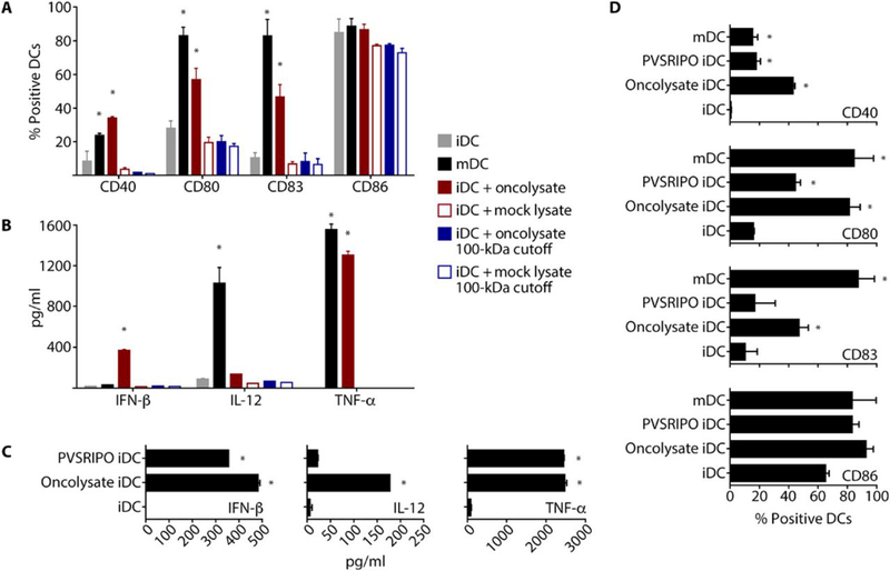
Fig. 3. PVSRIPO-induced oncolysate mediates DC activation.

Human DCs (immature DCs, iDCs; mature DCs, mDCs) were generated, and iDCs were treated as indicated. iDCs and mDCs were used as controls, as indicated in the figure. (A and B) Supernatant from untreated or PVSRIPO-infected (MOI=0.1; 48 hpi) DM6 melanoma cells was either unfiltered or filtered through a 100 kD cut-off filter. iDCs were treated with the resulting supernatants (24 h). Flow cytometry (A) and ELISA (B) were used to assess DC activation/maturation phenotype, viability, and pro-inflammatory cytokine production. (C and D) Human DCs were treated with DM6 oncolysate produced as in (A) or PVSRIPO at a titer equal to the amount detected in DM6 oncolysate. Supernatants were assessed for cytokine production by ELISA (C) and cell phenotype by flow cytometry (D). (A and D) For representative flow cytometry data see fig. S2. Experiments were repeated three times with cells from 2 donors; error bars denote SEM, and asterisks denote significant (p<0.05) ANOVA protected Tukey’s HSD test compared to mock controls.
