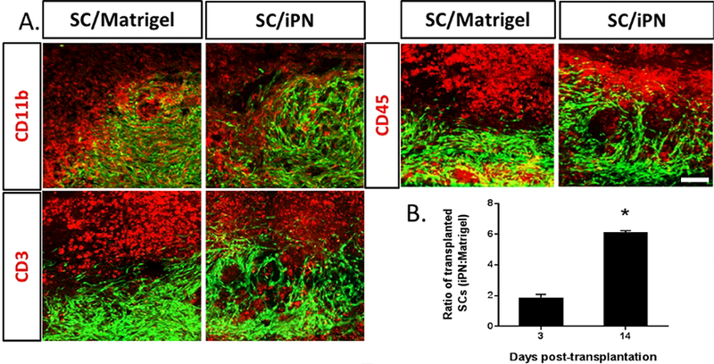
Figure 5.

A. Fluorescent micrographs of sagittal spinal cord sections at 14 days posttransplantation to evaluate immune cells. SCs (green) transplanted either in Matrigel or iPN were stained for microglia/ macrophages (CD11b), T cells (CD3) and leukocytes (CD45), in red. Bar = 100 μm. B. Ratio of transplanted SCs in iPN: transplanted SCs in Matrigel at 14 days are shown. Results expressed as mean ± SEM. * p<0.05
