
Figure 1. Effect of increased blood α2-antiplasmin levels on MMP-9 expression.
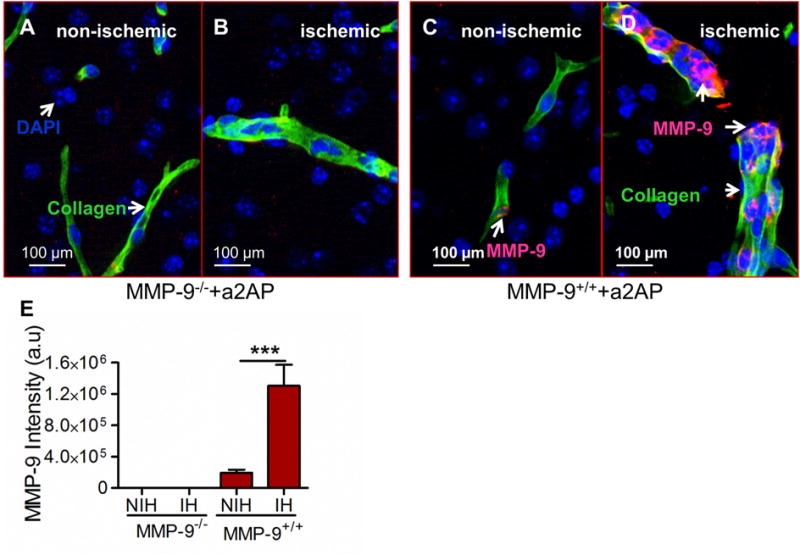
MMP-9+/+ and MMP-9−/− mice were intravenously supplemented with α2-antiplasmin prior to MCA thromboembolic stroke. Brain tissue was examined 24 h later. (A and B) Representative merged images (scale bar=100 μm) of the ischemic (IH) and non-ischemic (NIH) hemispheres after immunostaining for type IV collagen (green, blood vessels) and MMP-9 (red) in (A, B) MMP-9−/− mice and (C, D) MMP-9+/+ mice. Areas with both type IV collagen and MMP-9 stain yellow. MMP-9 expression (red color) inside type IV collagen (green, blood vessels) is indicated by white arrows. DAPI stained nuclei are blue in color. (E) Bar graph showing total MMP-9 intensity in arbitrary units (a.u.) in the ischemic hemisphere(IH) and non-ischemic hemisphere (NIH) of MMP-9+/+ mice. N= 4-6 ***p<0.001.